Learn About 4D Ultrasounds
A 4D ultrasound is an advanced imaging technique that provides real-time, three-dimensional views of the developing fetus, including its movements and activities. The "4D" refers to the three spatial dimensions (length, width, and depth) combined with the fourth dimension of time, which captures the motion of the fetus.


Bonding Experience:
One of the significant benefits of 4D ultrasounds is the enhanced bonding experience it offers to parents. Seeing the fetus's movements and facial expressions in real-time can strengthen the emotional connection between parents and their unborn child. It provides an opportunity to share in the joy and excitement of pregnancy.
Safety:
Similar to other ultrasound procedures, 4D ultrasounds are considered safe and non-invasive. They use sound waves instead of radiation, making them a preferred imaging modality during pregnancy. However, it's important to follow appropriate guidelines and recommendations provided by healthcare professionals.
Procedure:
The procedure for a 4D ultrasound is similar to that of a 2D or 3D ultrasound. A gel is applied to the mother's abdomen to facilitate sound wave transmission, and a transducer is used to emit high-frequency sound waves into the body. The sound waves then bounce back and are converted into detailed images using computer software, providing a real-time video-like display of the fetus's movements and anatomy.
Visualization:
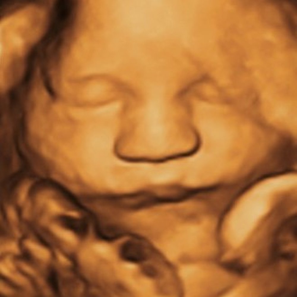
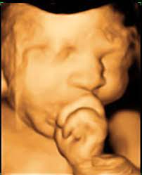
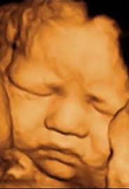
4D ultrasounds offer a more comprehensive and realistic representation of the fetus compared to traditional 2D or even 3D ultrasounds. Parents and healthcare providers can see the baby's facial features, expressions, gestures, and body movements in motion. It allows for a more immersive experience and emotional connection with the unborn child.
Image Quality
4D ultrasounds provide three-dimensional images of the fetus in motion, creating a realistic representation of the baby's features. The images are typically displayed in color and have depth, allowing for a more detailed visualization compared to the traditional 2D ultrasound.
Limitations:
While 4D ultrasounds provide impressive visualizations, they also have some limitations. The image quality may vary depending on factors such as fetal position, maternal body habitus, and the amount of amniotic fluid. In some cases, it may be challenging to obtain clear images due to factors beyond control.
